Pairwise interactions in nucleic acid 3D structures
Overview
The annotations of pairwise interactions listed below were developed for RNA, but apply equally well to DNA, except that in the absence of an O2' atom some interactions simply do not form. Additional perspectives can be found in the RNA 3D Structure Course that we developed.Annotations are calculated by the program NA_pairwise_interactions.py in the fr3d-python GitHub repository. As of 2026-03-04, the most recent version are in the "latest" branch.
Examples of all interactions can be found in the human ribosome; start at this link and select the different types of interactions with the drop-down in the tab header. Enter an interaction type in the filter box to narrow down the list.
Basepairs
We annotate RNA and DNA basepairs using the 12 basepairing families described by Leontis, Stombaugh, and Westhof (2002). Three regions of the bases are identified as "edges", the Watson-Crick edge, which participates in the most common A-U, A-T, and G-C basepairs, also the Hoogsteen edge and the Sugar edge. In all, there are 12 families of basepairs. Exemplar instances of each basepair in each family are shown at the RNA Basepair Catalog hosted by NAKB. This resource is highly recommended for understanding how the Leontis-Westhof system works, and how the Watson-Crick AU, AT, and GC basepairs fit into the cWW family, family #1.A few annotations go beyond Leontis, Stombaugh, and Westhof (2002). We follow the paper Westhof, Watson, Zirbel, Cate (2023) to annotate anionic GU and GG cWW basepairs; these are available in the basepair_detail tab and are annotated cWWa, where a stands for "anionic" or "alternative set of hydrogen bonds". We also annotate two different sets of hydrogen bond for UU, one called tWW, the other called tWWa. Also, we follow the 13th table in Leontis, Stombaugh, and Westhof (2002) by annotating some cis interactions where one base uses the Watson-Crick edge and one uses a bifurcated edge; these are annotated as cWB.
Base-phosphate interactions
Base-phosphate interactions are hydrogen bonding interactions between the three edges of nucleic acid bases (Watson-Crick, Hoogsteen, Sugar) and the phosphate oxygens of the same nucleotide or another nucleotide. They were described by Zirbel, Šponer, Šponer, Stombaugh, and Leontis (2009). Annotations are labeled 0BPh, 1BPh, ..., 9BPh; interactions that fall close to but outside the classification limits are annotated as "near" and indicated n0BPh, ..., n9BPh. See the 10 categories at the Base-Phosphate Catalog.Base-ribose interactions
Base-ribose interactions are hydrogen bonding interactions between the three edges of nucleic acid bases (Watson-Crick, Hoogsteen, Sugar) and the ribose oxygens (O2', O3', O4') of the same nucleotide or another nucleotide. They were described in the Appendix of Zirbel, Roll, Sweeney, Petrov, Pirrung, Leontis (2015). Annotations are labeled 0BR, 1BR, ..., 9BR; interactions that fall close to but outside the classification limits are annotated as "near" and indicated n0BR, ..., n9BR. Refer to the Base-Phosphate Catalog to see the locations of the hydrogen bonds around the bases, keeping in mind that the phosphate groups depicted there are replaced by ribose oxygens in base-ribose interactions.Base stacking interactions
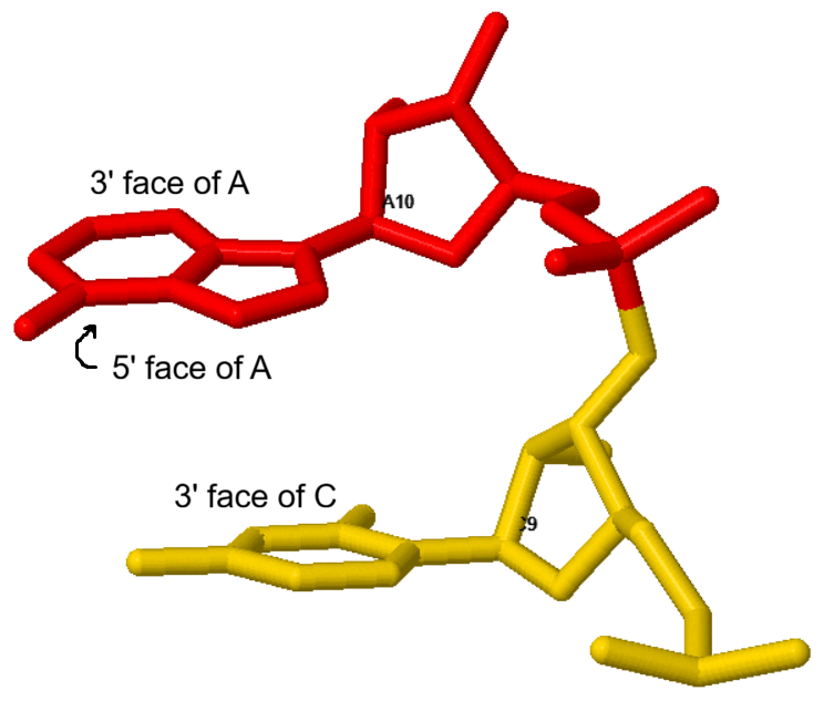
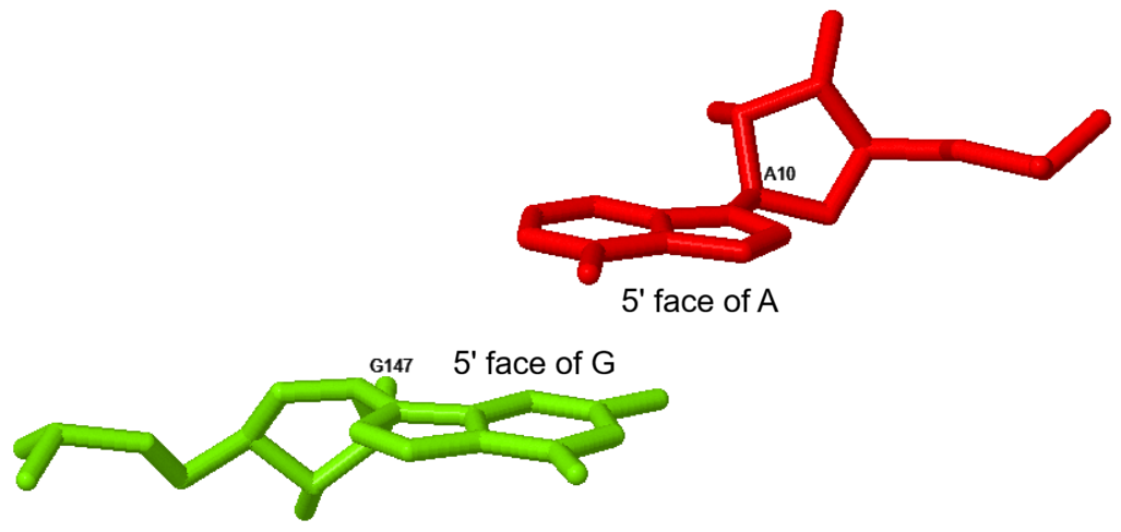
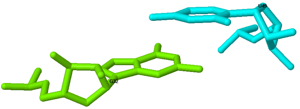
Base stacking occurs between adjacent bases in a double helix, but can also occur across the helix and in a variety of motifs. Base stacking was briefly described in the paper introducing the FR3D motif search tool, Sarver, Zirbel, Stombaugh, Mokdad, Leontis (2008). We name the two "faces" of each nucleotide base, the 3' face and the 5' face; when the 3' face of one base is stacked on the 5' face of another base, we call that s35 stacking. For example, in a regular double helix, a typical stacking interaction between successive bases would be reported as C9 s35 A10; the 3' face is toward the 3' end of the chain, and the 5' face is toward the 5' end. We could equivalently write the nucleotides in the other order and say A10 s53 C9. Cross-strand stacking is typically s55 stacking; the 5' faces make contact across the helix. There is also s33 stacking, most commonly in loop motifs. Here are examples of each distinct type of stacking from the human ribosome large subunit.- C9 s35 A10 ← follow the link to click and rotate the coordinates of the two nucleotides
- A10 s55 G147
- G32 s33 U49
Base oxygen stacking interactions
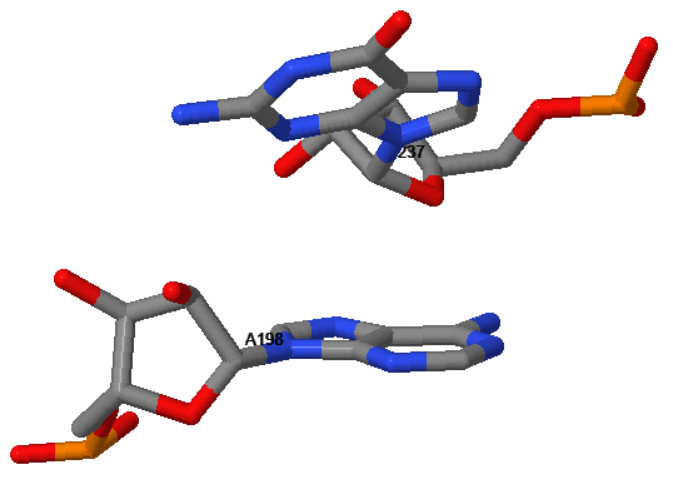
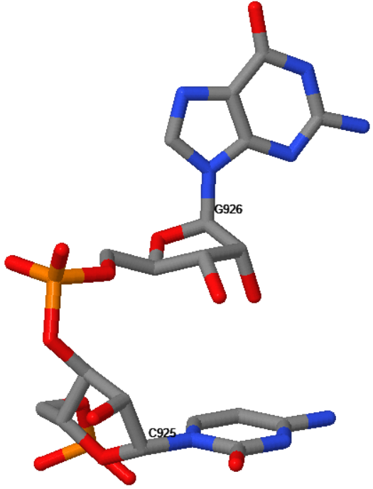
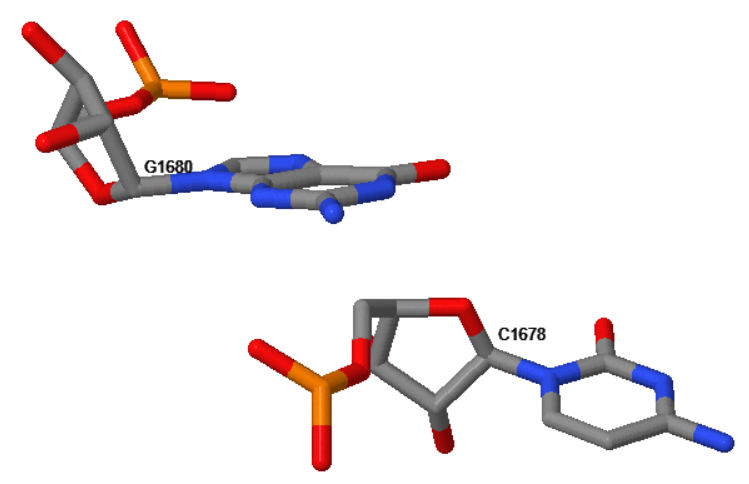
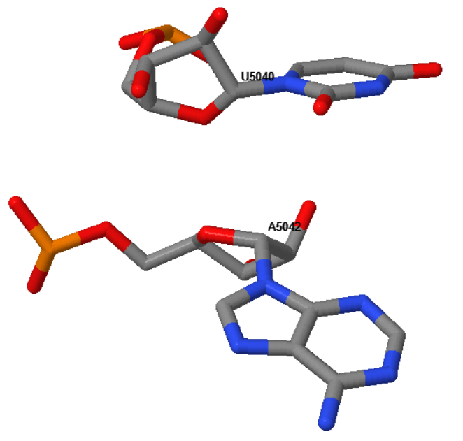
Many examples occur where the ribose or phosphate oxygen of one nucleotide stacks on the face of another nucleotide. These interactions were described in Zirbel and Auffinger (2022). We annotate the interactions with "s" for stacking, then "3" or "5" for the face, then one of the atoms from the list O2',O3',O4',O5',OP1,OP2. Here are some examples of possible annotations, with links to a coordinate viewer for each.- A198 s3O4' G237 Note that the 3' face of A is up, toward the O4' of G.
- C925 s3O3' G926
- G1680 s5OP1 C1678
- U5040 s5O2' A5042
Sugar-ribose interactions
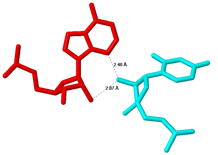
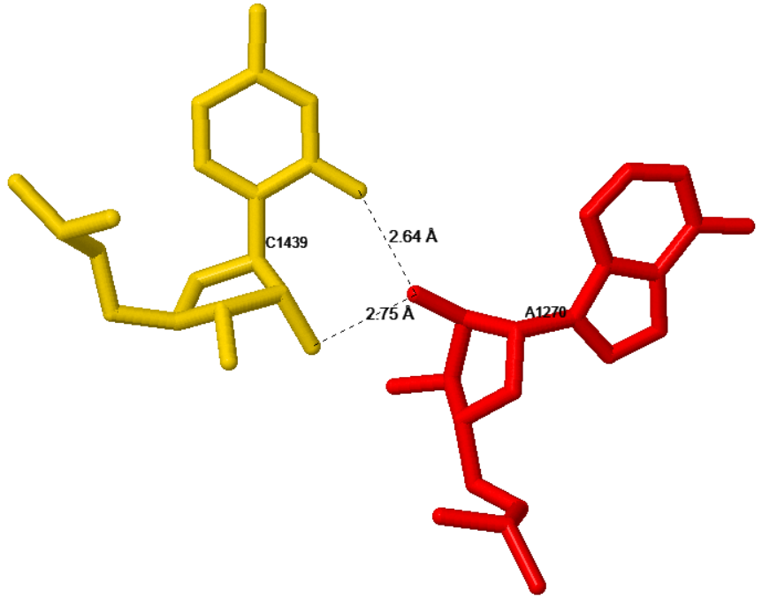
Sugar-ribose is a new annotation we developed for RNA-RNA interactions having simultaneous hydrogen bonds between the sugar edge and O2' atom of one nucleotide and the O2' atom of a second nucleotide. See the bonds marked in the images below. These are non-base-specific interactions; all four bases are able to make a hydrogen bond with their sugar edges; atoms are A(N3), C(O2), G(N3), U(O2). These contacts occur in the ribose zipper, in the A-minor interaction, and in other places.For a sugar-ribose annotation, the hydrogen bond lengths must both be shorter than 3.8A, and the O2' atom of the second nucleotide must be within 2A of the plane of the base of the first nucleotide. We annotate two different orientations, cis and trans, by analogy with the Leontis-Westhof basepair system. See the examples below.
- A62 cSR U77
This cSR interaction is also a cSS basepair; about 50% of cSR interactions are also cSS basepairs.
Note that the glycosidic bonds are on the same sides of the bases here.
- C1439 cSR A1270
This cSR interaction is not a cSS basepair; the bases are too far away for cSS.
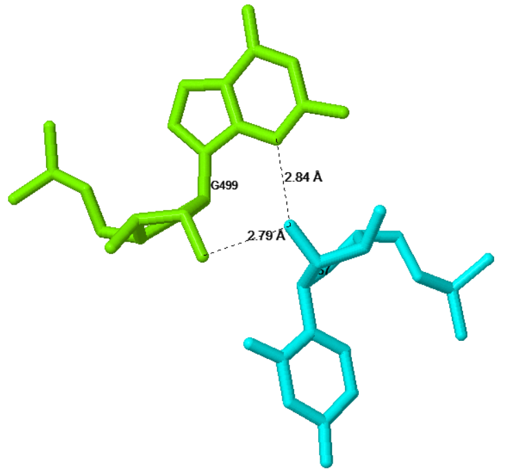
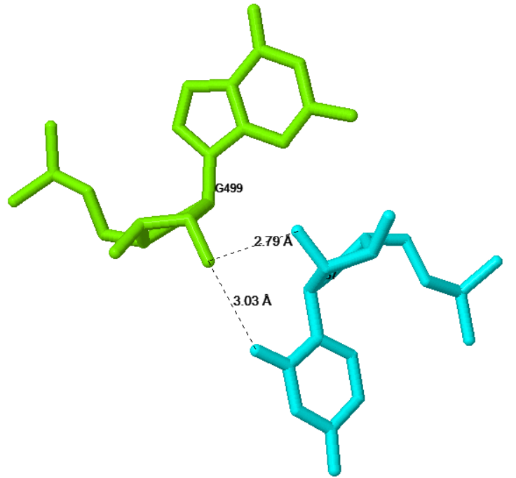
- G499 tSR U57
This tSR interaction shows the "trans" orientation of the glycosidic bonds; they are on opposite sides of the bases.
The hydrogen bond contacts are marked below left.
It is also the case that U57 makes a tSR interaction with G499; those hydrogen bonds are marked below right and would be annotated U57 tSR G499, or as G499 tRS U57.